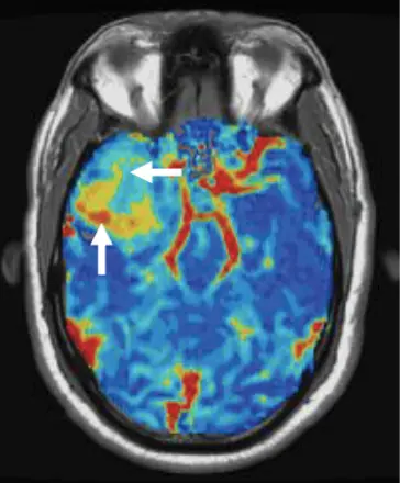
IB Neuro is an exclusive MRI perfusion platform that generates quantitative relative cerebral blood volume (rCBV) maps. The quantitative output has undergone independent validation with tissue samples and has been shown to far exceed the accuracy and repeatability over other perfusion methods. With over 30 years of pioneering development, this cutting-edge technology excels in differentiating tumor types, predicting tumor grades, and distinguishing post-treatment effects from tumor recurrence. It is the only platform proven reliable using 50% less gadolinium-based contrast agent for both 1.5T and 3T. Additionally, IB Neuro processes CT perfusion datasets, making it an indispensable tool in advanced neuroimaging.

IB Neuro automatically generates standardized sRCBV maps that quantify tumor vascularity with clarity and consistency—streamlining decision-making across neuro-oncology care.
What makes IB Neuro unique?
- First commercial DSC-MRI perfusion analysis platform1
- Incorporates the most-proven contrast leakage algorithm2
- Only DSC-MRI analysis software which uses patented standardization technology3
- Only DSC-MRI analysis software used in national multi-center clinical trials incorporating DSC-MRI4
- Independently validated with tissue samples5
- Direct comparison studies demonstrate superiority of IB Neuro over other platforms6
- Only platform PROVEN reliable using 50% less gadolinium contrast agent7
- Results in the GREATEST inter-reader agreement and confidence; levels the playing field based on user experience8
Support for superiority of IB Neuro
- FDA clearance received May 15, 2008 ↩︎
- IB Neuro incorporates the most-proven contrast leakage algorithm, which was co-developed by IB co-founder Dr. Kathleen Schmainda. Dr. Schmainda has performed numerous studies to validate the IB Neuro contrast agent leakage method and implementation. IB Neuro is the only platform to incorporate this version – proven with over 20 years of studies. One example ↩︎
- IB Neuro is the only DSC-MRI analysis software that uses patented standardization technology.
First paper describing standardized rCBV with improved consistency:
Bedekar D, Jensen T, Schmainda KM. Standardization of relative cerebral blood volume (rCBV) image maps for ease of both inter- and intrapatient comparisons
Standardized rCBV demonstrates improved repeatability compared to normalized methods available on other platforms:
Prah MA, Stufflebeam SM, Paulson ES, Kalpathy-Cramer J, Gerstner ER, Batchelor TT, Barboriak DP, Rosen BR, Schmainda KM. Repeatability of Standardized and Normalized Relative CBV in Patients with Newly Diagnosed Glioblastoma ↩︎ - IB Neuro is the only DSC-MRI analysis software used in national multi-center clinical trials which incorporate DSC-MRI. Study results show that IB Neuro-generated rCBV can be used to predict outcomes. This also demonstrates the ability of IB to reliably analyze data from all MRI vendor platforms.
ACRIN 6677
ACRIN 6684
ACRIN 6686
ECOG-ACRIN (EAF151 – active clinical trial)
GABLE (EAF223 – active clinical trial) ↩︎ - IB Neuro-generated rCBV has been independently validated with tissue samples:
Demonstrated ability of IB Neuro to estimate tumor burden
Demonstrated ability of IB Neuro rCBV to distinguish tumor from treatment effect ↩︎ - Direct comparison studies demonstrate the superiority of IB Neuro over other platforms:
IB Neuro proved more accurate than NordicICE ↩︎ - IB Neuro is the only platform PROVEN reliable using 50% less gadolinium contrast agent:
Using a low flip angle acquisition and IB Neuro post-processing, 50% less contrast can be used with the result comparable to the standard double-dose method ↩︎ - IB Neuro’s standardized rCBV and FTB maps result in the GREATEST inter-reader agreement and confidence, and level the playing field based on user experience.
Arterial Spin-Labeling and DSC Perfusion Metrics Improve Agreement in Neuroradiologists’ Clinical Interpretations of Posttreatment High-Grade Glioma Surveillance MR Imaging − An Institutional Experience ↩︎