Neuro-Oncology Solutions
When it comes to brain tumor imaging, analysis software applications are NOT all created equal. Colorized functional maps may appear similar at first glance, but a closer look reveals critical differences in how they are calculated and their underlying meaning. Backed by over 30 years of focused research in Neuro-Oncology, Imaging Biometrics’s applications are trusted by leading brain tumor centers to provide imaging insights that help guide their most important decisions. IB sets the standard with a complete suite of applications for analyzing advanced MR images.
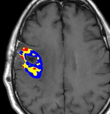
IB sets the standard with a complete suite of applications for analyzing advanced MR images. IB’s applications enhance standard anatomical imaging with key physiological information like true regions of contrast enhancement (void of post-surgical blood products), and tumor blood volume and flow, helping radiologists, surgeons and oncologists make better decisions for their patients. For patients who cannot undergo MRI, IB’s applications are also compatible with CT images. IB’s applications have been proven reliable and accurate in recent ground-breaking multi-center studies1,2 and are the most trusted by clinical trials researchers for their imaging needs3,4.

1 Impact of Software Modeling on the Accuracy of Perfusion MRI in Glioma
Hu LS, Kelm Z, Korfiatis P, et al.
AJNR Am J Neuroradiol. 2015 Dec;36(12):2242-9. doi: 10.3174/ajnr.A4451.
2 Performance of Standardized Relative CBV for Quantifying Regional Histologic Tumor Burden in Recurrent High-Grade Glioma: Comparison Against Normalized Relative CBV Using Image-Localized Stereotactic Biopsies
Hoxworth JM, Eschbacher JM, et al.
AJNR Am J Neuroradiol. 2020 Mar;41(3):408-415. doi: 10.3174/ajnr.A6486.
3 Quantitative Delta T1 (dT1) as a Replacement for Adjudicated Central Reader Analysis of Contrast-Enhancing Tumor Burden: A Subanalysis of the American College of Radiology Imaging Network 6677/Radiation Therapy Oncology Group 0625 Multicenter Brain Tumor Trial
Schmainda KM, Prah MA, et al.
AJNR Am J Neuroradiol. 2019 Jul;40(7):1132-1139. doi: 10.3174/ajnr.A6110.
4 ACRIN 6684: Multicenter, phase II assessment of tumor hypoxia in newly diagnosed glioblastoma using magnetic resonance spectroscopy
Ratai EM, Zhang Z, et al.
PLoS One. 2018 Jun 14;13(6):e0198548. doi: 10.1371/journal.pone.0198548.